FMD Studio
Flow Mediated Dilation software reliable endothelial function assessment
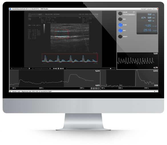
Real-time Analysis
Automatic Doppler Flow Analysis
High Accuracy
ECG Gating Not Required
Quality Feedback
It is clinically challenging to evaluate the Flow Mediated Dilation without appropriate instrumentation.
Our software together with your ultrasound system, can automatically measure
the diameter of the vessel and calculate the percentage of FMD.
Features
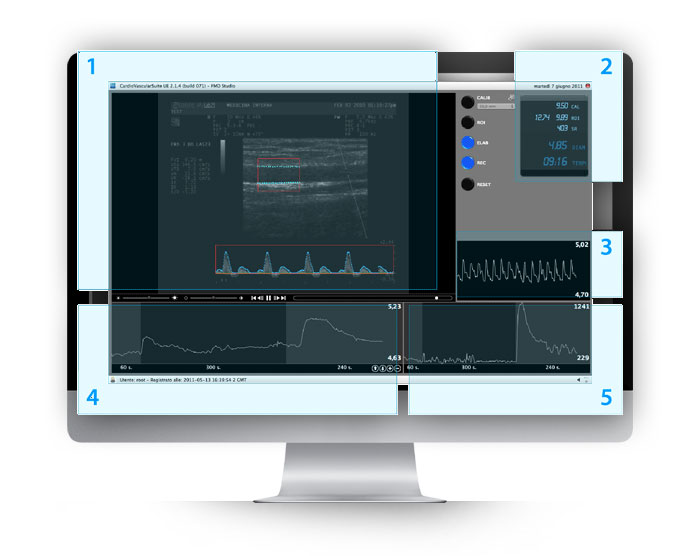
Real-time Analysis
This is a great advantage in studies of FMD, where one of the most critical points is to maintain good image quality for the duration of the test (9-10 minutes). With the aid of a real-time system, the sonographer can more easily adjust the position of the ultrasound probe to compensate for the movements of the patient.
Automatic Doppler Flow Analysis
In addition to the automatic measurement of the diameter, the software automatically analyzes the Doppler signal (when available in dual-mode by ultrasound equipment) in order to calculate the value of instantaneous shear rate. This provides the exact amount of the vasodilation stimulus, which can be used to normalize the FMD value, as strongly recommended by more recent studies.
High Accuracy
The system uses an innovative mathematical operator with sub-pixel precision. This permits to overcome the spatial resolution limitations of the image with results otherwise obtainable only through a more complex analysis of the ultrasound radio frequency signal.
ECG Gating Not Required
ECG gating is not required. This type of measurement usually requires synchronization with the ECG. The technique we developed overcomes this requirement in favor of the simplicity of the procedure and the cost of the required equipment.
Quality Feedback
The system provides signals that can be used by the sonographer as markers of quality and reliability of the images.
User Interface

Endothelial Function and Atherosclerosis
The endothelium plays a central role in the initiation, progression and clinical outcomes of atherosclerosis. Flow-mediated dilation (FMD) of the brachial artery is an established non-invasive method used to assess endothelial function. The method was introduced by Celermajer et al. in 1992, and over the last 20 years has become increasingly important since several studies have shown that an impaired FMD response is related to cardiovascular risk factors such as smoking, hypercholesterolemia, hypertension, diabetes and aging, and is an independent predictor of cardiovascular events. Indeed, thousands of papers have established that endothelial dysfunction is one of the earliest detectable signs of atherosclerosis in patients ranging from youngsters to adults. FMD can be measured by ultrasound imaging, as described in the guidelines of the International Brachial Artery Reactivity Task Force (Corretti et al., 2002). The examination consists in measuring brachial artery diameter at rest and after reactive hyperemia induced by ischemia of the forearm. The measurement is made on a B-mode section of the artery, which is imaged above the antecubital fossa in the longitudinal plane.

Validation
FMD Studio has been validated in 50 healthy subjects (mean age 25 ± 5 years) and 220 patients with more than one cardiovascular risk factor (mean age 59 ± 14 years). Flow-mediated dilatation was lower in patients than in healthy subjects (5.3 ± 2.9% vs 7.1 ± 2.5%, p = 0.001), while mean brachial diameter at rest was significantly larger in patients (4.32 ± 0.09 mm vs 4.19 ± 0.08 mm, p = 0.01). The system has also been validated in a multicenter study involving seven centers and 675 FMD/GTN scans. Overall intra-day variability (1 hour apart) was CV = 9.9%, overall inter-day variability (1 month apart) was CV = 12.9%. The results showed that FMD Studio provides reproducible FMD assessment and can be adopted in multicenter clinical trials.
FMD Studio is now used in clinics worldwide.
Equipments
Our probe holder especially designed for FMD studies. The video acquisition hardware devices that you can use with Cardiovascular Suite.
User manual
Cardiovascular Suite 4 User Manual.
A printed copy of the User Manual and Instruction for Use can be requested at support@quipu.eu
Video tutorial
See how to install, configure and use Cardiovascular Suite.
Brochure
We invite you to download a 14-day fully functional trial version of Cardiovascular Suite, including FMD Studio and Carotid Studio. The software runs on Apple and Windows Computers, activation by email and Internet connection are required.
Minimum system requirements:
APPLE COMPUTER
Apple Mac Computer with: Intel Core i5 5th generation 2.3 GHz Turbo boost, 4GB RAM, 1GB free Hard Disk space*, USB 3.0 port, 1280x800 monitor resolution, Mac OS X 10.12 - 10.15.
MICROSOFT WINDOWS COMPUTER
Intel Core i5 5th generation 2.3 GHz Turbo boost, 4GB RAM, 1GB free Hard Disk space*, USB 3.0 port, 1024x768 monitor resolution.
Microsoft Windows 7 64 bit, Windows 8.1 64 bit, Windows 10 64 bit, OpenGL ES 2.1.
(*) 250GB free Hard Disk space is suggested for the Archive
Suggested computer:
APPLE COMPUTER
MacBook Air o MacBook Pro, processor M2 or later, 8-16GB RAM, 512GB SSD (larger for more than 1000 FMD studies), Display 13’’-16’’, macOS 13 or superior.
MICROSOFT WINDOWS COMPUTER
Laptop with Intel Core i5-i7 12000+ or AMD Ryzen 5-7 5000+, 16GB RAM, 512GB SSD (larger for more than 1000 FMD studies), Display 14’’-16’’, resolution 1920x1080, Windows 10 or 11.

非常抱歉,您只有购买软件后才能查看完整软件教程!
